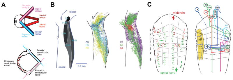
Figure 4.
Organization of the vestibular system. A. Schematics depicting the spatial arrangement of the six eye muscles (top) and the three semicircular canals of the ipsilateral labyrinth (bottom). Color-coded antagonistic pairs of eye muscles are spatially aligned with individual semicircular canals. B. Spatial arrangement of the vestibular nuclei on a horizontal section through the dorsal hindbrain (left) and color-coded overlay of afferent projections from the three semicircular canals (middle) and the three otolith organs (right). C. Schematic depicting the rhombomeric arrangement of spinal projecting (green) and midbrain oculomotor nucleus projecting (red) neurons (left) in the amphibian. Right: Allocation of classical vestibular nuclei nomenclature (yellow) onto the amphibian rhombomeric scaffold and tentative segmental location of vestibular subgroups that mediate sensory signals from specific semicircular canals onto spatially matching extraocular and spinal motoneuronal populations. ACe/ACi/PCe/PCi, excitatory/inhibitory neurons of anterior/posterior semicircular canal; ATD, ascending tract of Deiters; DVN/LVN/MVN/SVN, descending/lateral/medial/superior vestibular nucleus; HCe/HCi, horizontal semicircular canal excitatory/inhibitory neurons; INT, abducens internuclear neurons; IO/IR, inferior oblique/rectus motoneurons; MR/LR, medial/lateral rectus motoneurons; LVST, lateral vestibulo-spinal tract; r1–r8, rhombomeres 1–8; SO/SR, superior oblique/rectus motoneurons; Tan, tangential nucleus; VN, vestibular nuclei. B and C are adapted with permission from [Straka et al. 2014; Straka et al. 2002], respectively.

