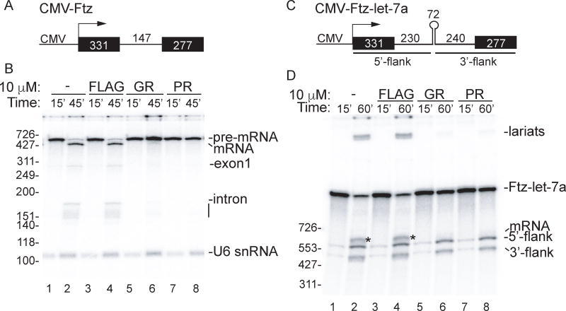
Figure 1. GR and PR toxic peptides inhibit splicing in vitro.
(A) Schematic of CMV-Ftz DNA template used for transcription-coupled splicing showing the CMV promoter and sizes of the exons and intron. (B) CMV-Ftz DNA was incubated in transcription-coupled splicing reaction mixtures for 15 minutes with no peptide or in the presence of 10 µM GR, PR or FLAG peptides. α-Amanitin was then added to stop further transcription, and incubation was continued for 30 minutes to allow splicing. RNA species are indicated. The line below the intron marks intron breakdown products. (C) Schematic of CMV-Ftz-let-7a DNA template which contains a pri-let-7a sequence in the Ftz intron. (D) CMV-Ftz DNA was incubated in transcription/splicing/pri-miRNA processing reaction mixtures for 15 minutes with 10 µM of the indicated peptides. After adding α-Amanitin, incubation was continued for 45 minutes. The asterisks on the gel indicates the spliced mRNA. Markers in nts are shown. See also Figure S1.

