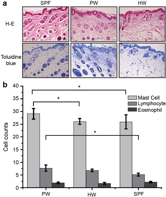
Figure 2.

Infiltration of eosinophils, mast cells, and lymphocytes in AD skin lesions. a) Representative images of skin tissue sections were examined after H&E staining (lymphocytes and eosinophils) or toluidine blue staining (mast cells). b) Immune cells were counted under a microscope and the results expressed as the mean±SEM of the number of cells per 0.025 mm2 of skin at week 4 (n=4 mice per group); *p<0.05. Red arrowheads indicate mast cells in toluidine blue staining. Blue arrowheads indicate lymphocytes. Black arrowheads indicate neutrophils. AD - atopic dermatitis, HW - hydrogen water, SPF - specific-pathogen-free, PW - purified water
