
Fig. 1.
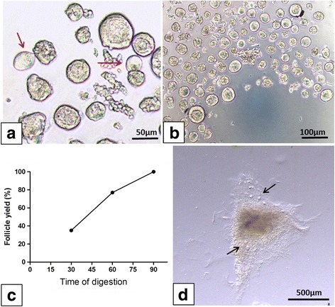
Isolation of human preantral follicles. In the previous protocol, some follicles showed extruded oocytes (red arrow) (a); in the modified protocol, the vast majority of follicles were isolated from ovarian tissue samples (b). Distribution of isolated follicles in the modified protocol according to the three time intervals (c); some follicles remained entrapped in fragments of undigested tissue (black arrows) (d)