
Fig. 2.
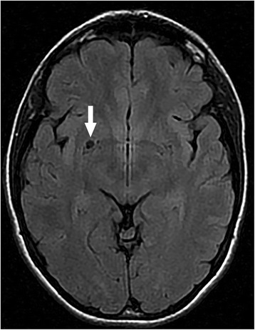
Magnetic resonance imaging (MRI) scan. Dilated atypical Virchow-Robin space (VRS) under the basal ganglia on the right side is visible (white arrow)
Official websites use .gov
A
.gov website belongs to an official
government organization in the United States.
Secure .gov websites use HTTPS
A lock (
) or https:// means you've safely
connected to the .gov website. Share sensitive
information only on official, secure websites.
Magnetic resonance imaging (MRI) scan. Dilated atypical Virchow-Robin space (VRS) under the basal ganglia on the right side is visible (white arrow)