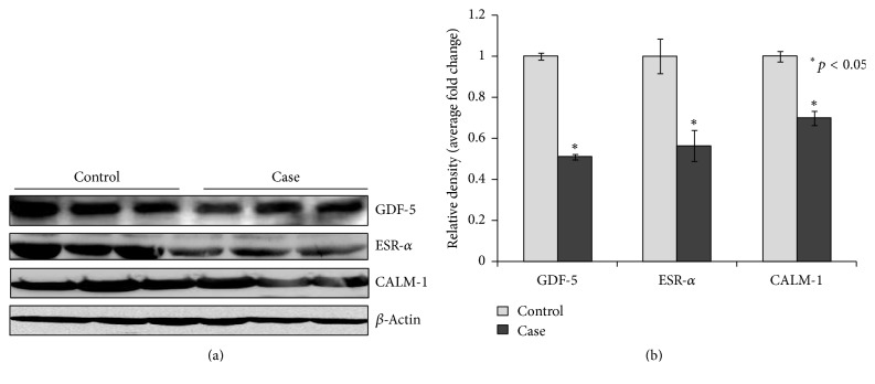
Figure 3.
(a) Western blot analysis was performed to understand the protein level of GDF5, ESR-α, and CALM-1 in control and case study. (b) Bar diagram showing the relative protein density after normalization with β-actin. Relative protein density of GDF5, ESR-α, and CALM-1 was significantly decreased in case study as compared to control group. Representative blots showing three samples from each group (control and case). Values are expressed as mean ± SEM (n = 24). ∗p < 0.05 versus control.

