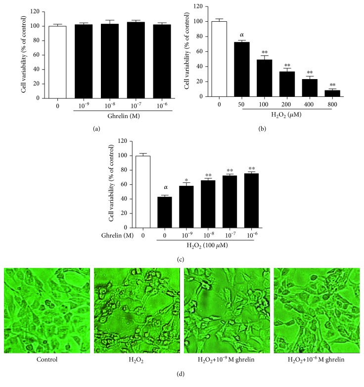
Figure 1.
The effects of ghrelin on the cell viability of H2O2-treated HLECs. (a) HLECs were incubated with different concentrations of ghrelin (10−9–10−6 M) for 24 h. (b) HLECs were incubated with different concentrations of H2O2 (50–800 μM) for 24 h. (c) HLECs were preincubated with ghrelin (10−9–10−6 M) for 12 h before being treated with 100 μM H2O2 for 24 h. Cell viability was assessed via MTT assay. (d) HLECs were detected by microscopy. The results were represented as the mean ± SEM (n = 3) from three independent experiments. αP < 0.01, compared with the untreated control group; ∗P < 0.05, ∗∗P < 0.01, compared with the H2O2-treated group.

