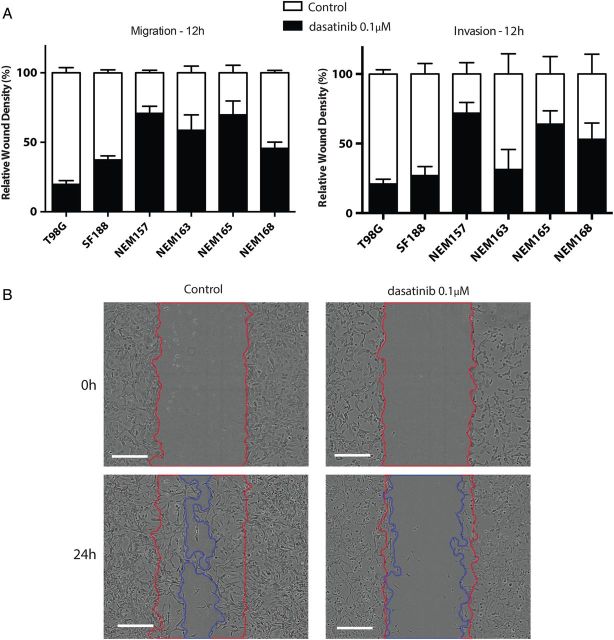
Fig. 3.
Effect of dasatinib on cell migration and cell invasion. Cells were seeded in 96-well plates and incubated until subconfluent. A wound was scratched across each well (WoundMaker, Essen BioScience), and BD Matrigel was then added (or not for migration assay) with treatment. Wound closure was automatically imaged each 6 h and calculated as a percentage of wound confluence that the cell gained. (A) Quantitative wound-repair analysis at 12 h after dasatinib treatment (0.1 μM) for migration and invasion assay, respectively. (B) Contrast-phase images of NEM168 invasion at 24 h. Red and blue lines correspond to the frontline of the scratch wound at 0 h and 24 h, respectively. Scale bar = 300 µM.

