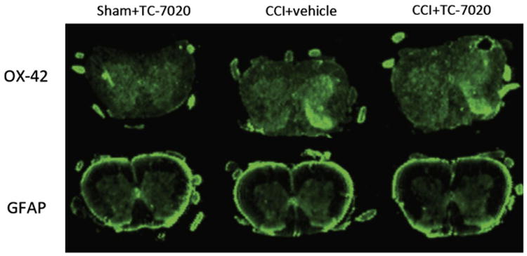
Figure 3.

Example of the immunohistochemistry of OX42 and glial fibrillary acid protein, markers of microglial and astrocyte activation respectively, of the L4-6 spinal cord in CCI+vehicle, CCI+3 mg/kg/day of TC-7020 and sham+3 mg/kg/day TC-7020. The laminae I and II of the ipsilateral dorsal horn show increased staining in rats with CCI compared to sham-operated rats.
