
FIG. 7.—
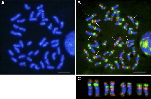
Fluorescence in situ hybridization of centromeric and telomeric repeats. (A) DAPI banding of metaphase chromosomes. Chromosome preparation was obtained from peripheral blood leucocytes of fugu. (B) Two-color FISH using the 118-mer centromeric and the telomeric (TTAGGG)n repeats probes. The centromeric and telomeric probes are labeled in green and red, respectively. Arrows indicate interstitial telomere-like sequence (ITS). (C) Confirmation of the ITS localization. These signals are shown on four chromosome pairs of fugu. Scale bar represents 5 μm.