FIGURE 1.

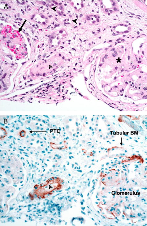
Representative renal biopsy specimen from a subject with histologic evidence of TA-TMA after hematopoietic stem cell transplantation. A, hematoxylin–eosin staining (magnification, ×20) of renal cortex with glomeruli. Glomeruli show variable degrees of obliteration of the capillary lumens and thickened capillary walls (asterisk) with mild to moderate mesangial matrix expansion. There is also evidence of red blood cell fragmentation and extravasation (arrow). There are no inflammatory infiltrates in glomeruli. Small arterioles (A) also show obliteration of the vessel lumen due to sloughed endothelial cells, intimal proliferation, and extracellular matrix deposition. There is mild tubular atrophy as well as mild interstitial inflammation (arrowheads). B, C4d staining (magnification, ×20) of corresponding tissue section shows diffuse positive staining in the degenerating small arteriole (A) with microangiopathic changes. Focal or rare C4d stain is noticeable in some tubular basal membranes (Tubular BM), PTC, and glomeruli. PTC, peritubular capillary; TA-TMA, transplant-associated thrombotic microangiopathy.
