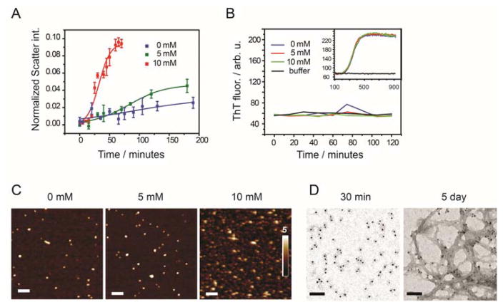
Figure 1.
(A) Scatter intensity of freshly prepared monomeric Aβ-42 (concentration 10 μM) incubated at 25°C in PBSN buffer with 0 mM glucose (blue), 5 mM glucose (green) and 10 mM glucose (red) at pH 6.8 obtained from dynamic light scattering. Scatter signals of buffer without Aβ was subtracted and intensities were normalized to toluene scatter signals; means ± SD, n = 3. (B) ThT fluorescence of 10 μM freshly filtered Aβ-42 allowed to aggregate in the DLS cuvette under the same conditions as in (A). Samples were collected at different time point, diluted 3-fold into same buffer + ThT (20μM). Within 2 hours of incubation no ThT positive signal was found. After 2 h the diluted sample was further incubated at 37˚C (inset) and ThT-positive aggregates were formed. (C) AFM images of 10 μM freshly filtered Aβ-42 incubated for 15 minutes in PBSN buffer containing 0 mM, 5 mM, and 10 mM glucose at pH 6.8. Scale bars 100 nm. (D) Immuno TEM images of 5 μM Aβ-42 incubated in GS buffer of pH 6.8 after 30 minutes and after 5 day incubation; Scale bars 100 nm.

