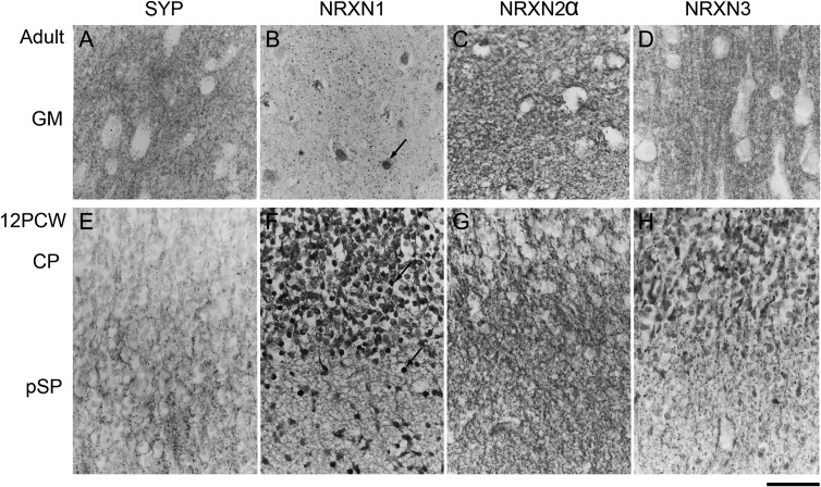
Figure 2.
Immunohistochemistry for SYP and the NRXNs in paraffin sections from adult (A–D) and fetal (E–H) cerebral cortex. (A) Punctate SYP immunoreactivity confined to presumptive synaptic terminals in the gray matter neuropil. In the fetal cortex (E) such punctate staining is largely confined to the pSP. All three NRXNs also exhibited punctate staining (B–D) in adult cortex, although at a lower density for NRXN1 with evidence of nuclear staining (arrow, B). In the fetal cortex, NRXN1 was expressed in cell bodies, processes, and some nuclei (arrows) of many immunoreactive cells in the pSP and CP but without punctate staining (F). NRXN2, however, exhibited punctate staining in the pSP in particular (G). NRXN3 was expressed in cell bodies and processes mostly in the CP, with some punctate immunoreactivity, largely in the pSP (H). Scale bar = 50 µm.

