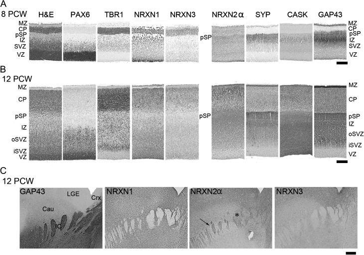
Figure 3.
A comparison of NRXN immunoreactivity with other cell markers across the cortical wall at 8 PCW (A) and 12 PCW (B). H&E, hemotoxylin and eosin stained; o, outer; i, inner. Note that PAX6 revealed radial glial progenitor cells; TBR1, postmitotic neurons; SYP, CASK and GAP43 neurites of the pSP and IZ. NRXN1 and 3 predominantly localized to layers with a high-cellular density, whereas only NRXN2α predominantly colocalized with synaptogenic zones and growing axons. (C) Internal capsule in the ventral forebrain in which bundles of growing axons were GAP43 positive, NRXN1 and 3 negative, and partially positive for NRXN2α (arrow) although some axon bundles appeared negative (*). Scale bars: A and B, 200 µm; C, 500 µm.

