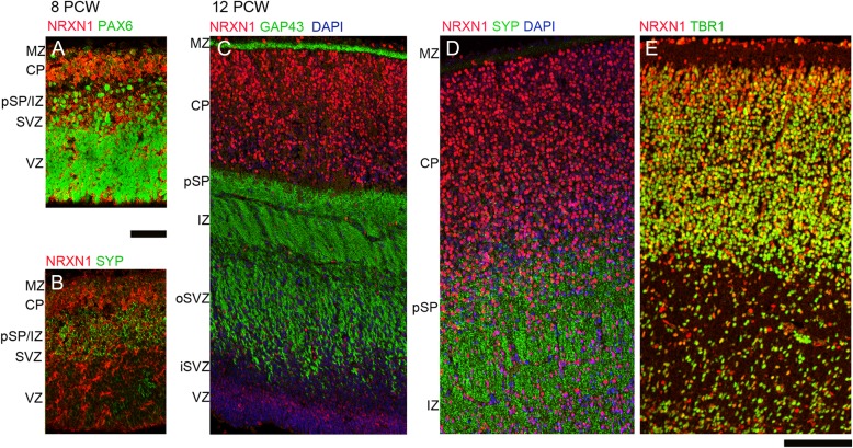
Figure 4.
Colocalization of NRXN1 immunoreactivity (red) with phenotypic markers (green) by immunofluorescence. At 8 PCW NRXN1 immunoreactivity was present in cytoplasm/membranes around PAX6-positive radial glia, particularly near the apical ventricular surface (A) but showed little colocalization with SYP in the pSP at either 8 PCW (B) or 12 PCW (D) nor with GAP43 in growing axons of the IZ (C). However, there was strong colocalization (orange–yellow) with TBR1 in the postmitotic neurons of the CP. Scale bars = 100 µm.

