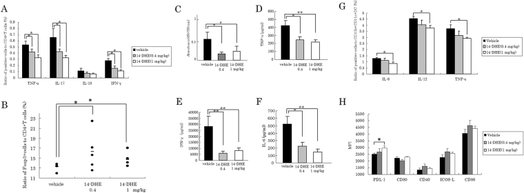
Figure 6.
14-DHE suppressed the production of inflammatory cytokines and cell expansion, and increased Tregs in LN T cells. (A and B) To detect cytokine-producing cells and Tregs, LN cells from mice immunized with MOG/CFA were cultured under stimulation with Leukocyte Activation Cocktail plus BD GolgiPlug, and analyzed by flow cytometry. (A) Ratios of TNF-α-, IL-17-, IL-10- and IFN-γ-producing cells among CD3e and CD4-positive cells. (B) Ratios of CD3e, CD4, CD25 and Foxp3-positive cells among CD3e and CD4-positive cells. (C–F) To detect cell proliferation and cytokine production, LN cells were cultured with 2 μM MOG. BrdU was added to the cell culture to detect cell proliferation. (C) Concentrations of pro-inflammatory cytokines produced in response to re-stimulation with MOG antigen. (D,E, and F) ELISA quantification of TNF-α, IFN-γ, and IL-6, respectively, in the cell supernatant. (G) To detect cytokine production in mDCs, LN cells were stimulated with Leukocyte Activation Cocktail plus BD GolgiPlug, and the ratio of IL-6-, IL-12p70-, and TNF-α positive cells was measured among CD11b and CD11c-positive cells by flow cytometry. (H) Expression of the cell-surface markers PDL-1, CD80, CD40, ICOS-L and CD86 on CD11b and CD11c-positive myeloid DCs was analyzed by flow cytometry.

