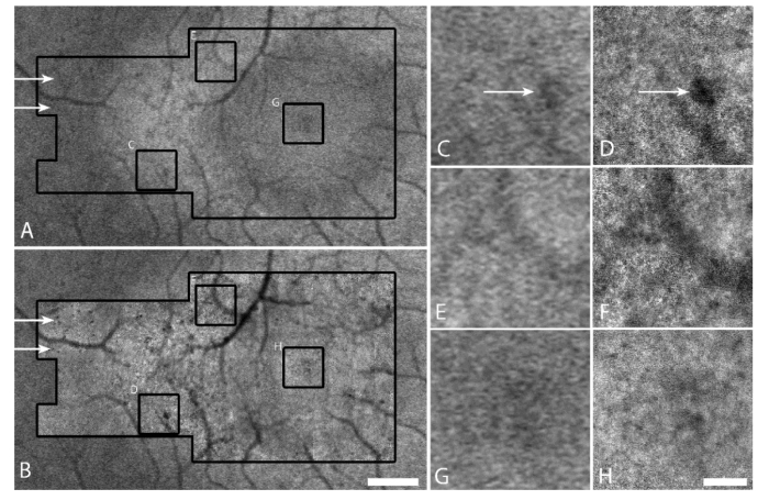
Fig. 1.
Comparison of conventional IRAF to AO-IRAF for a patient with retinitis pigmentosa. (A) Conventional IRAF image acquired using a Heidelberg Spectralis. Black outline shows the location where AO-IRAF images were taken. (B) AO-IRAF montage (inside larger black outline) overlaid on the conventional IRAF image. Scale bar, 0.5 mm. (C-H) Zooms of three areas outlined by the small black squares in (B) comparing conventional IRAF (C,E,G) to AO-IRAF (D,F,H). Scale bar, 100 µm. The RPE mosaic is visible in (D) and (F) in some portions of the image. Additional details about hypofluorescent areas can be observed using AO-IRAF (arrows). The hyperfluorescent ring in (A) is less visible in (B) due to the stretching of each individual image within the AO-IRAF montage, an artifact of montaging.

