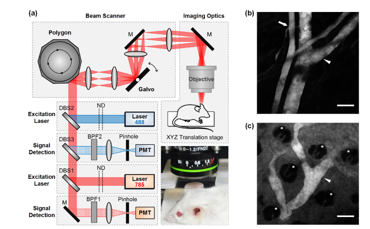
Fig. 1.
(a) Schematic of laser-scanning confocal microscopy: DBS, dichroic beam splitter; BPF, band pass filter; ND, neutral density filter; M, mirror; PMT, photomultiplier tube. Photograph shows an anesthetized live mouse prepared for ear skin imaging, in vivo. (b, c) The representative images acquired at the ear skin of mouse after the intravenous injection of ICG. (b) At immediately after the ICG injection, blood vessels were visible. The artery (arrow) and vein (arrow head) were distinguishable by the vessel diameter and blood flow speed. (c) At 30 minutes after the ICG injection, the lymphatic vessel (arrow head) draining ICG leaked from the blood vessel to dermal tissue was visible. The hair follicle (asterisk) was also distinguishable. Scale bar: 100 μm.

