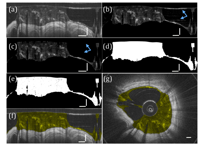
Fig. 4.
Segmentation of airway mucus. (a) The input image. (b) The image after filtering and thresholding are performed. (c) Morphological operations are performed to remove artifacts (blue arrows). (d) The resulting binary mask, (e) applied to image (b). The segmented mucus is color coded yellow in (f) polar and (g) Cartesian coordinates. a-f) vertical scale bars, 0.5 mm; horizontal scale bars, 30°. (g) scale bar, 0.5 mm.

