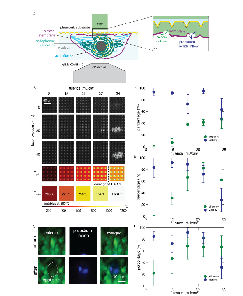
Fig. 1.
Using plasmonic substrates to investigate plasma membrane poration of differentiating myoblasts with fluorescent imaging.(A)The experimental setup consists of an 850-ps laser source illuminating a plasmonic pyramidal substrate consisting of uniform base lengths (2.4 μm), heights (1.4 μm), and base to base spacings (1.2 μm) with adherent C2C12 cells. The substrate-cell composite is placed upside down on a glass coverslip with 200 µL of phosphate-buffered saline containing propidium iodide. The assembly is placed on an objective for fluorescence imaging. The inset shows laser-induced poration, enabling the exchange of intra- and extracellular molecules. (B) Shows bright-field images of the pyramidal substrate exposed at different laser fluences and exposure times to determine the optical damage threshold of the plasmonic substrate. At the bottom are single laser shot simulation results of the temperature of the gold pyramids and surrounding water. (C) Cells pre-stained with Calcein AM confirm plasma membrane poration. A decrease in calcein signal after laser exposure indicates outflow of intracellular molecules, while an increase in propidium iodide signal indicates inflow of extracellular molecules. The poration efficiency of C2C12 as a function of fluence for different laser exposures: (D)10 ms, (E) 20 ms, and (F) 40 ms. Data show standard error of the mean for three independent measurements with around 3-6 cells in each spot.

