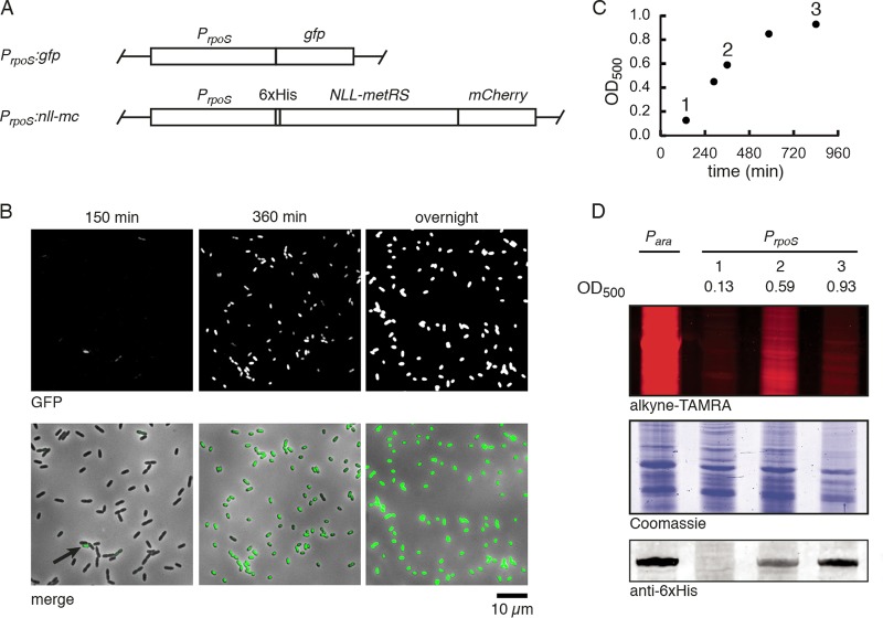
FIG 1 .
Cell-state-selective labeling using the rpoS promoter. (A) P. aeruginosa was engineered to express GFP or an NLL-MetRS–mCherry translational fusion under control of the endogenous rpoS promoter. Expression cassettes were transposed to the Tn7 chromosomal locus. (B) Representative images of GFP fluorescence of the PrpoS:gfp strain throughout growth in LB medium. GFP fluorescence (top) and a GFP–bright-field merge (bottom) are shown. The arrow indicates a GFP-positive cell at the early time point. The times after 1:200 dilution into fresh medium are indicated above the panels. (C) Optical density at 500 nm of PrpoS:nll-mc cells grown in liquid culture in minimal FAB medium. At each indicated time point, an aliquot was removed and incubated with 1 mM Anl for 15 min. (D) Lysates were treated with alkyne-TAMRA and separated via SDS-PAGE to visualize Anl incorporation. Coomassie staining of the same gel indicates equal protein loading. Lysates were probed by Western blotting for the six-histidine tag on NLL-MetRS.

