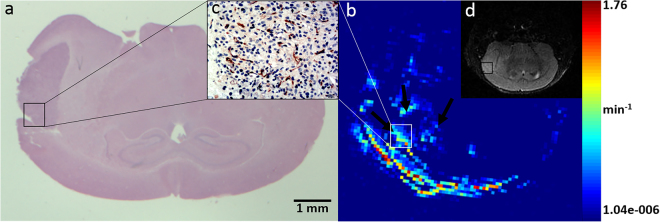
Figure 1.
MRI and corresponding histopathological features of the orthotopic U87MG glioblastoma model. HE staining (a) and a T2-weighted image (d) show a quasi-circular mass in the right cerebral hemisphere with mild edema around the tumor mass. The Ktrans map (b) of the tumor is shown. Bright points in the tumor area represent high permeability, especially in the tumor margin, which also showed more abundant neovascularization (c). All images were selected from the maximum cross-section of the tumor.

