Figure 3.
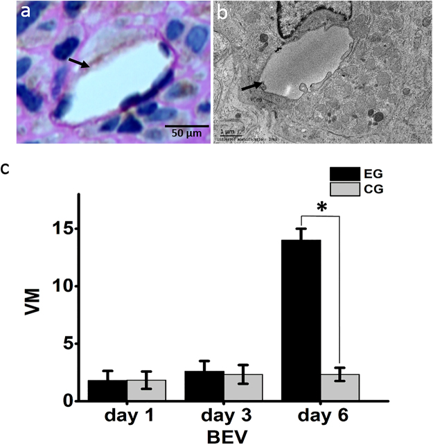
Vascular mimicry. (a) Vascular mimicry was characterized by PAS-positive and CD34-negative lumen-like structures (black arrow). (b) Transmission electron microscopy shows tumor cells lining a vascular channel (black arrow). (c) The amount of vascular mimicry (VM) was significantly increased 6 days after BEV administration, experimental group (EG), control group (CG). Data were represented as the means ± SD. *Indicates P < 0.05.

