
Figure 4.
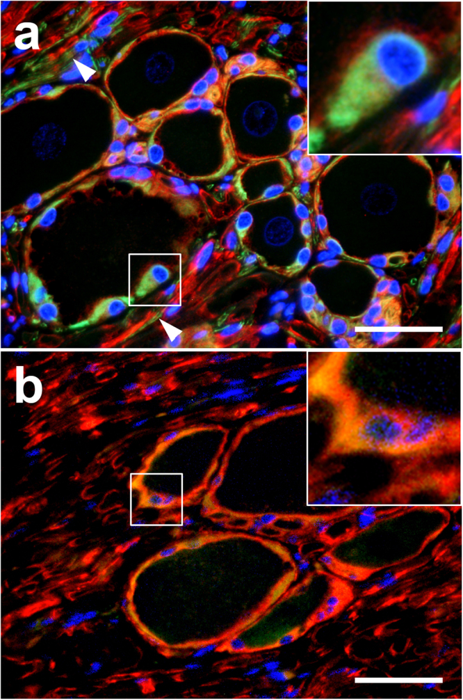
Dorsal root ganglion of a Beagle dog. (a) 2′,3′-cyclic-nucleotide 3′-phosphodiesterase (CNPase; red) and glial fibrillary acidic protein (GFAP; green) double-staining. Most satellite glial cells (SGCs) have a strong perinuclear CNPase and cytoplasmic GFAP expression (insert). Note CNPase+ nerve fibers surrounded by GFAP+ cells (arrowheads). (b) CNPase (red) and Nestin (green) double-staining. Most SGCs show a strong co-expression of these markers indicated by yellow staining (insert). Immunofluorescence double-labelling of the dorsal root ganglion of a beagle dog in situ with bisbenzimide as nuclear counterstain. Bar, 40 μm.