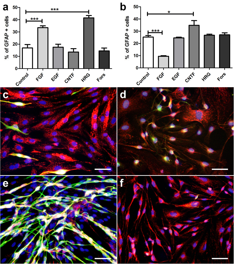
Figure 8.
GFAP expression of canine (a,c,e) and murine (b,d,f) satellite glial cells (SGCs) supplemented with fibroblast growth factor 2 (FGF), epidermal growth factor, (EGF), ciliary neurotrophic factor (CNTF), heregulin 1β (HRG), and forskolin (Fors). (a,b) Graphs show the percentage of glial fibrillary acidic protein (GFAP)+ SGCs with and without supplementation. (c–f) Immunofluorescence double-labelling of SGCs for 2′,3′-cyclic-nucleotide 3′-phosphodiesterase (CNPase; red) and GFAP (green) with bisbenzimide as nuclear counterstain. (c,d) control medium. (e,f) FGF supplementation. *P < 0.05. ***P < 0.001. Bars, 60 μm. Shown are means with standard errors of the mean (SEM).

