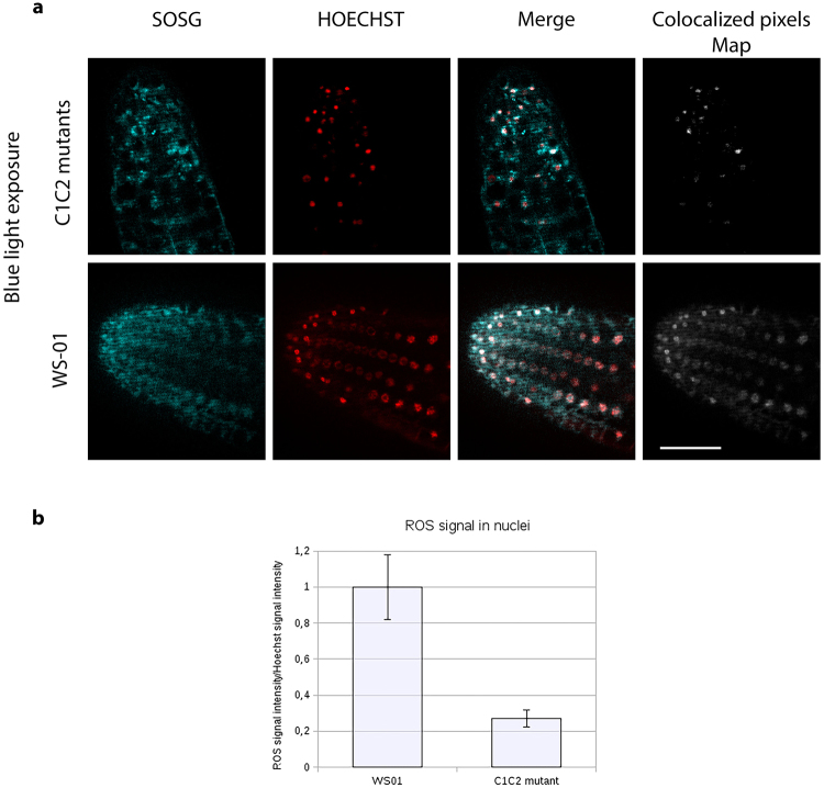
Figure 2.
Production and subcellular localization of ROS in Cryptochrome-1 overexpressing (WS01) and double mutant cry1cry2 (C1C2) seedlings exposed to blue light. Primary root tips from 8-day old etiolated Arabidopsis seedling (see methods) roots either lacking both cry1 and cry2 (C1C2 mutant) or over- expressing cryptochrome 1 (WS01) were treated with HOECHST for nuclear staining and SOSG for ROS staining for 30 minutes, exposed to blue light for 10 minutes, and then immediately viewed by a Zeiss AxioImager.Z1/ApoTome microscope. (a) Images show single z section that cross the nucleus. Colocalized pixels appear in white on the merged images and on the colocalized pixels maps. Scale bars are 25 μm. (b) using the segmentation and ROI manager tool on imageJ, fluorescence intensity of white and red pixels were quantified from these images for each nucleus and exported to LibreOffice calculator. The mean fluorescence intensity of >50 individual nuclei is shown.

