Figure 4.
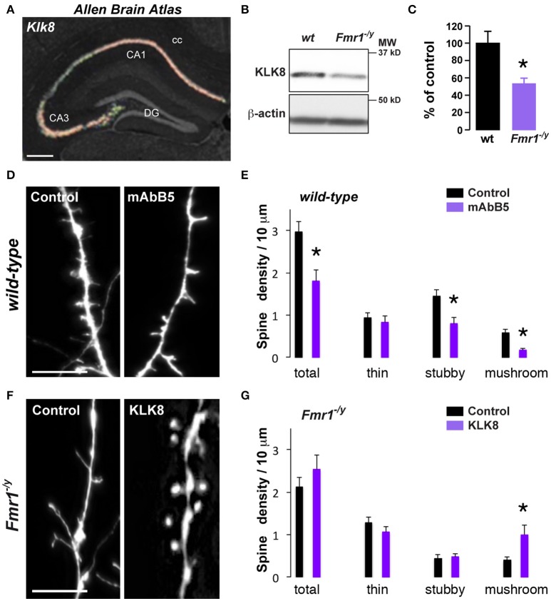
Decreased Klk8 expression in Fmr1−/y mice alters hippocampal dendritic spine maturation. (A) ISH coronal section from Allen Brain Atlas showing the enrichment of Klk8 in the dorsal hippocampus. (B) WB of KLK8 in hippocampal homogenates of wild-type (wt) and Fmr1−/y mice. (C) Quantification of KLK8 expression expressed as percentage of KLK8 expression in wild-type (wt) mice (n = 6 mice/genotype). (D) Red fluorescence from wt mice hippocampal primary culture (DIV14) incubated (mAbB5) or not (control, μg/35 mm dishes) with the activity-neutralizing anti-KLK8 antibody. Scale bar: 10 μm. (E) Quantification of spines density. (F) Red fluorescence from Fmr1−/y mice hippocampal primary culture (DIV14) transfected with DsRed alone (control) or DsRed and KLK8-Venus (KLK8). Scale bar: 10 μm. (G) Quantification of spine density in Fmr1−/y hippocampal neurons transfected or not with KLK8. Spine density (E,G) was quantified on three dendritic areas/neuron, 3–5 neurons/primary culture, and reproduced at least in 3 independent experiments per condition. DG, dentate gyrus; cc, corpus callosum. *p < 0.05.

