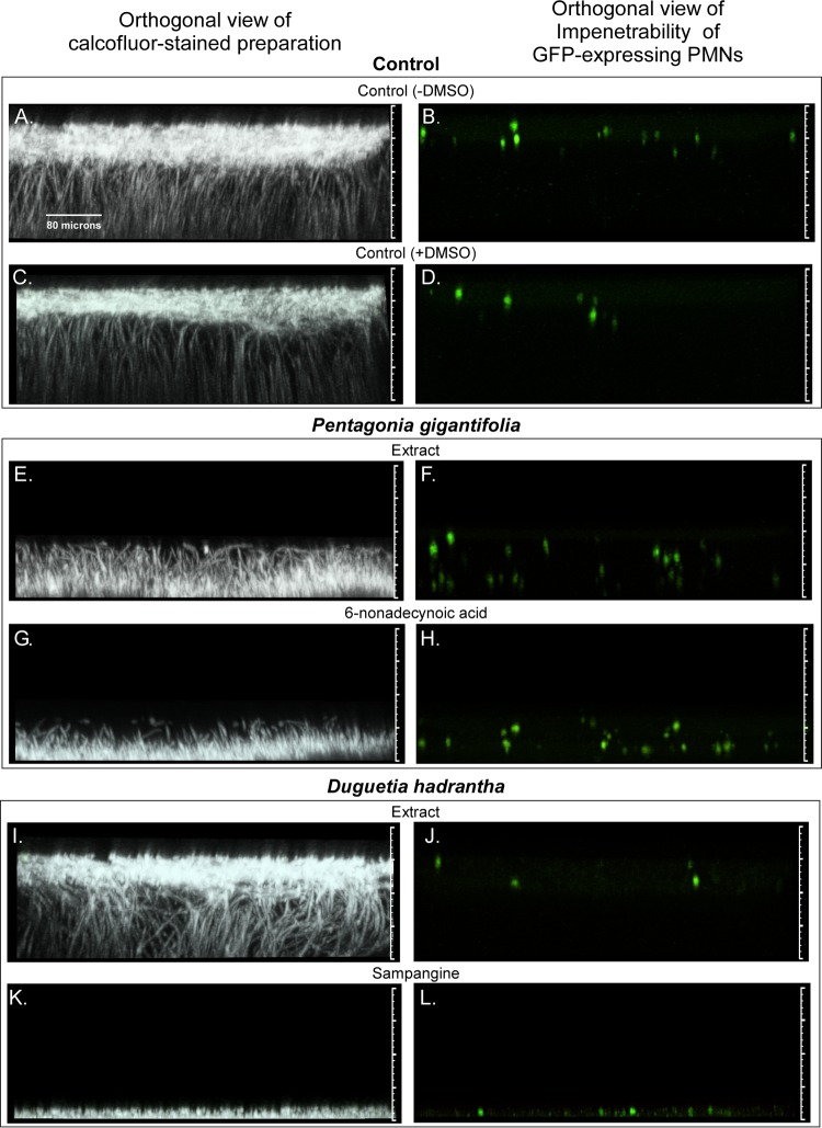
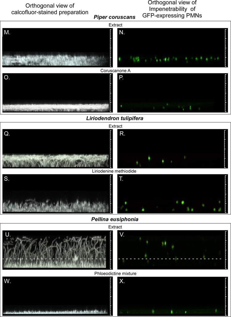
FIG 3.
Orthogonal (side) views of biofilm staining with calcofluor white or imaging of green fluorescent protein (GFP)-expressing PMNs after treatment with five pairs of extracts and derivative compounds previously shown to inhibit the growth of C. albicans. The five pairs of extracts and derivative agents are described in Table 1, and biofilm characteristics are described in Table 2. (A to D) Untreated biofilm control in the absence of DMSO (A, B) and untreated biofilm control in the presence of 0.4% DMSO (C, D). Note that all test biofilms treated with extracts or derivative agents contained 0.4% DMSO. (E to H) Representative biofilms treated with the extract of Pentagonia gigantifolia (E, F) or the derivative 6-nonadecynoic acid (G, H). (I to L) Representative biofilms treated with the extract of Duguetia hadrantha (I and J) or the derivative sampangine (K and L). (M to P) Representative biofilms treated with the extract of Piper coruscans (M, N) or the derivative coruscanone A (O, P). (Q to T) Representative biofilms treated with the extract of Liriodendron tulipifera (Q, R) or the derivative liriodenine methiodide (S, T). (U to X) Representative biofilms treated with the extract of Pellina eusiphonia (U, V) or the derivative phloeodictine mixture (W, X). The matted hyphae in the Pellina eusiphonia extract-treated preparation unwound when inverted, causing the biofilm, which was devoid of ECM, to appear thicker than it was. The dashed lines in panels U and V reflect the actual upright thickness in this case. The orthogonal views represent vertical slices (sections) through the stack of CLSM scans (projected image) that had been rotated 90°. The view is of an orthogonal slice taken 40 pixels into the projected image.


