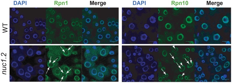
Figure 2.
Localization of 26S proteasome subunits in WT and nuc1.2 plants. Immuno-localization of Rpn1a (Left) and Rpn10 (Right) proteasome protein subunits in WT and nuc1.2 root tip cells. Arrows point foci of Rpn1a and Rpn10 proteins in nucleoli of nuc1.2 mutant cells. DAPI staining was used to visualize nucleoplasm and distinguish the nucleolus.

