Figure 4.
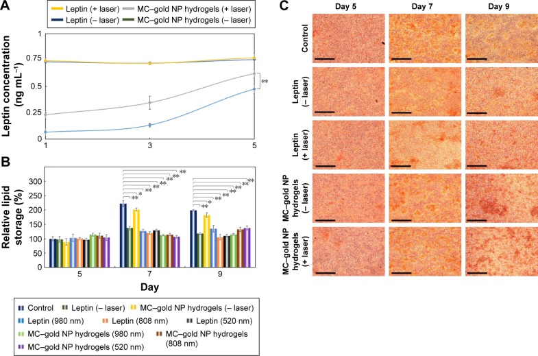
Light-triggered release of leptin from leptin-embedded MC–gold NP hydrogels.
Notes: (A) Measurement of the leptin release. Leptin-embedded MC–gold NP hydrogels were placed in Transwell™ inserts (Corning, New York, NY, USA) and incubated in complete DMEM at 37°C for irradiation (980 nm; 10 min) every day for 5 days. Leptin release was determined by Mouse Leptin ELISA Kit (Sigma-Aldrich Co., St Louis, MO, USA; **P<0.05; based on a two-tailed t-test, assuming unequal variances). The bars represent the mean ± standard deviation (n=4). Lines are included to guide the eye. (B) Quantification of lipid storage in cells was measured by the optical density (OD) of Oil Red O dye post-incubation under various conditions of light irradiation (*P>0.15; **P<0.05; based on a two-tailed t-test, assuming unequal variances). The bars represent the mean ± standard deviation (n=3). (C) Photographs of Oil Red O dye-stained cells at different periods post-treatment. Bars =200 µm.
Abbreviations: −, without; +, with; MC, methylcellulose; NP, nanoparticle.

