
Figure 2. Structural basis for binding of RNAP to the 30S subunit.
(A). Two binding sites of RNAP (boxed). (B) Close-up view of the Zn-finger interactions with S2. (C) Close-up view of the β−flap helix packing on S18. Molecular surface, secondary structure and sticks are shown in panels (B) and (C). (D) Position of two OB domains of S1 (orange) near the 3′ end of 16S rRNA (red). (E) Comparison of segmented maps of the 30S•RNAP complex formed with and without S1.

Figure 2—figure supplement 1. Crosslinked sites mapped on the 30S•RNAP structure.
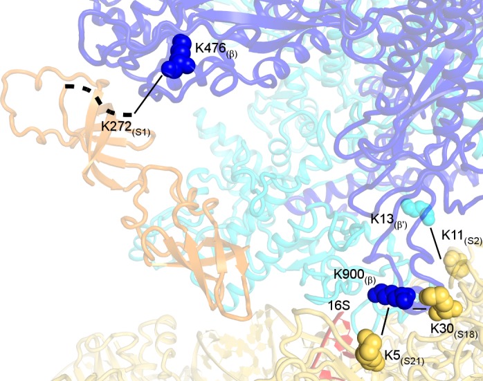
Crosslinks with e-value <1.0 × 10⁻³ and S1(K272)-β(K476) with e-value = 6.47 × 10⁻³ are shown.
Figure 2—figure supplement 2. Conformational rearrangements of RNAP between the non-rotated (blue) and rotated (gray) states.
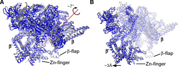
(A) Rotation of RNAP is shown relative to the 30S subunit (30S subunits were aligned). (B) A shift of the β′ subunit relative to the β-subunit (β-subunits were aligned).