. 2017 Oct 24;6:e29430. doi: 10.7554/eLife.29430

Figure 1. Two main functional conformations of BiP.

Ribbon representation of the structure of the ATP-bound BiP (PDB 5e84 [(Yang et al., 2015]), referred as the domain-docked conformation, and ADP-bound, which was modeled from the structures of the isolated NBD (PDB 5evz [Hughes et al., 2016]) and (D) SBD (PDB 5e85 [Yang et al., 2015]) and referred as the domain-undocked (U) conformation. The NBD (blue), βSBD (green), and αLid are highlighted by different colors and labeled.

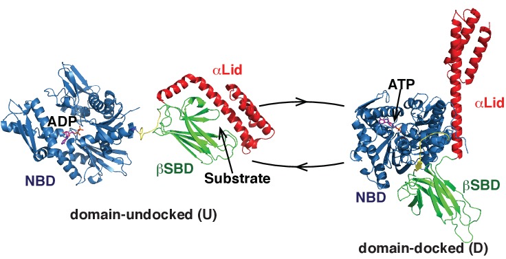
Figure 1.

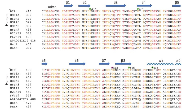
Figure 1—figure supplement 1. Sequence alignment of eukaryotic and bacterial Hsp70 proteins.

Figure 1—figure supplement 1.

The aligned sequences of the βSBD, including human HSP70s: BiP (GRP78 or HSPA5), HSP70 (HSPA1A or HSP72), HSPA2, HSC70 (HSPA8 or HSP73), HSPA9 (GRP75, Mortalin-2 or MTHSP70), and HSPA6; Hsp70s from eukaryotes: Stratiomys singularior (UniProt: B2CKI9), Plasmodiophora brassicae (UniProt: A0A0G4IKJ2) and Monodelphis domestica (UniProt: F6VP59); and bacterial Hsp70s: HscA and DnaK from Escherichia coli. Secondary structure elements are indicated above the protein sequence. The highly conserved residues are coloured in red, less conserved in orange, non-conserved in black. The positions of ‘soft mutations’ used in this study are indicated by green asterisks; the AMPylation site (Thr 518) is highlighted by a magenta asterisk.