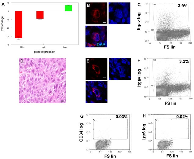
Figure 7. A new tumor initiating cell population with limited differentiation capacity from SCCs with EMT phenotype.

A. Increased expression of integrin αV (Itgav) in cells from SCC with EMT phenotype. qRT-PCR shows decreased expression of known cancer stem cell markers CD34 and Lgr6 in Itgav+ cancer cells. Experiments were performed three times with similar results. Error bars indicate SEM. B. Itgav+ cancer cell (red) in SCC with EMT phenotype. Nuclei were counterstained with DAPI. Scale bar = 5 μm. A representative section is shown. C. Flow cytometric sorting of Itgav+ cancer cells from SCC with EMT phenotype. Itgav log and forward scatter linear (FS lin) scales are shown. A representative sort is shown. D. H&E stained section of poorly differentiated SCC arising from transplanted Itgav+ cancer cells. E. Itgav+ cancer cell (red) in transplanted SCC. Scale bar = 5 μm. F. Flow cytometric sorting of Itgav+ cancer cells from transplanted SCC. Itgav+ cancer cells fail to regenerate CD34+ or Lgr6+ cancer stem cells. CD34+ G. and Lgr6+ H. cancer stem cells were sorted from transplanted SCC by flow cytometry.
