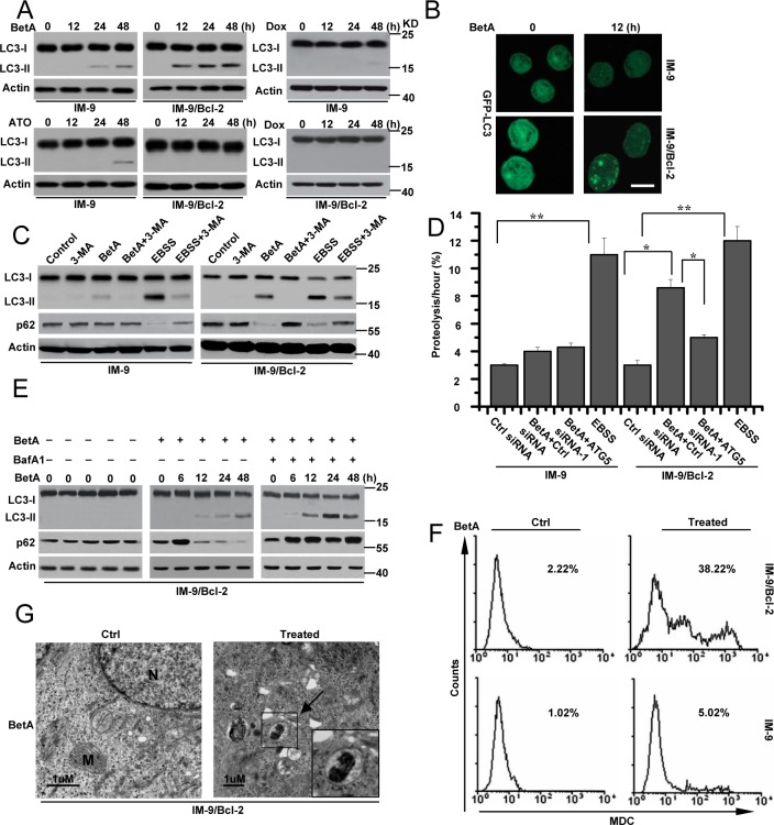
Figure 2. BetA induces autophagic cell death in IM-9/Bcl-2 not in IM-9 cells.
(A) immunoblot analysis of LC3-I and LC3-II levels in cells. Cells were treated as described in Figure 1, and then treated cells were collected for detecting LC3-I and LC3-II content by Western blotting, with β-Actin serving as a loading control. (B) Cells were treated as described in A. IM-9/Bcl-2/GFP-LC33 cell lines were treated with BetA for 12 h, fixed, and then visualized by fluorescent microscopy, Bars, 10 μM. (C) Autophagosome inhibitor 3-MA attenuated the effect of BetA on autophagy in IM-9/Bcl-2 cells. Cells were treated with BetA for 48 h in the absence or presence of the inhibitor of class III PI3 kinases 3-MA (5 mM), lysed and subjected to western blotting with anti-LC3 or p62 antibodies to monitor autophagy. EBSS, (starvation medium) is as a control, which is a classical stimulus used to induce the build-up of autophagosomes and autophagic flux. (D) BetA promotes long-lived protein degradation in cells cultured in full medium. IM-9 and IM-9/Bcl-2 cells were first transfected with Ctrl or ATG5 siRNA-1 for 24 h, and then radiolabeled for 24 h with 0.05 mCi/ml of L-[U-14C]valine. At the end of the labeling period, the cells were rinsed three times with phosphate-buffered saline. The cells were then incubated in full medium in the presence or in the absence of 10 μg/ml BetA or EBSS with 10 mM valine for 12 h. The data are presented as the means ± S.D. from three independent experiments (*,P<0.05; **,P<0.01). (E) IM-9/Bcl-2 cells were treated with BetA, and then 20 nM of BafA1 was added to the culture medium 2 h before cell lysis. Cells were treated with BetA at indicated time in the absence or presence of BafA1. p62 expression and LC3 conversion were detected by immunoblot analysis, with β-Actin serving as a loading control. (F) Indicated cell lines were treated with BetA for 48 h, stained with MDC and analyzed by flow cytometry. (G) Representative electron micrographs of IM-9/Bcl-2 treated or untreated with 10 μg/ml BetA for 24 h. Arrows, presence of autophagosomes. N, nucleus; M, mitochondria. Bars, 1μM. All data are representative of three independent experiments.

