
Figure 6. Lunasin decreases the level of PCSK9 secreted in hepatic tissues in ApoE−/− mice by immunofluorescence analysis.
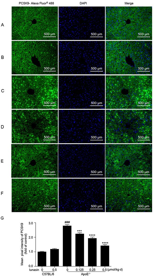
Hepatic tissue of mice was fixed in 4% paraformaldehyde and paraffin-embedded 4 μm sections subjected to immunofluorescence analysis. Representative images of liver paraffin sections stained for PCSK9 with Alexa Fluor® 488 (green) and counterstained with DAPI to show cell nucleus (blue). (A) C57BL/6 + vehicle (normal control); (B) C57BL/6 + 0.5 μmol/kg·d lunasin; (C) ApoE−/− + vehicle (model control); (D) ApoE−/− + 0.125 μmol/kg·d lunasin; (E) ApoE−/− + 0.25 μmol/kg·d lunasin; (F) ApoE−/− + 0.5μmol/kg·d lunasin. (G) Quantitative analysis of PCSK9 image pixel intensity. #### p < 0.0001 vs. C57BL/6 group; *** p < 0.001 and **** p < 0.0001 vs. ApoE−/−group. Data represented as the means ±SEM. The photomicrographs were taken by Zeiss AX10 fluorescence microcopy at original magnification × 200. Results are representative of three independent experiments with similar results.