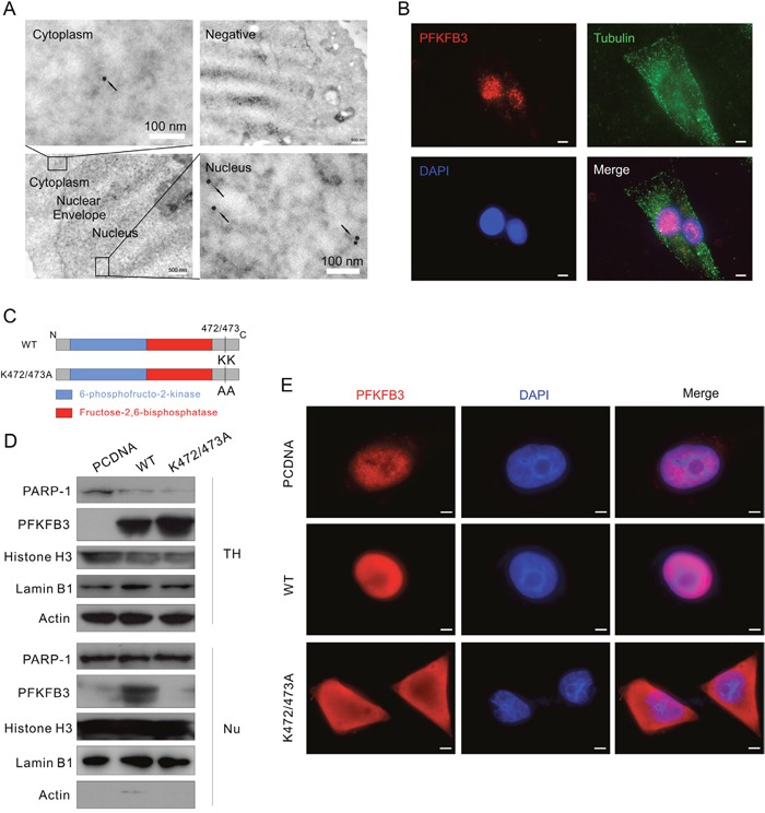
Figure 3. K472/473A mutation changes the nuclear localization of PFKFB3.

(A) HeLa cells were analyzed by immunoelectron microscopy using anti-PFKFB3 antibody. Images are representative of at least 20 cells. Arrowheads, PFKFB3 signals (gold particles). The negative control was prepared without addition of specific antibody. (B) Immunofluorescence was performed for ACHN cells using the antibodies of PFKFB3 and Tubulin, and the nuclei were stained by DAPI. Bar = 20 μm. (C) Schematic diagram of WT PFKFB3 (top) and K472/473A mutated PFKFB3 (bottom). (D and E) After transfection with the PCDNA, WT or K472/473A plasmids, TH and Nu subcellular fractions were extracted from ACHN cells and analyzed by immunoblotting with the antibodies indicated (D); cells were stained using the PFKFB3 antibodies and the nuclei were stained by DAPI (E). Bar = 10 μm. Images were representative of at least 20 cells.
