
Figure 4. PLGA NPs reduced Aβ40 and Aβ42 levels in AD mouse brains.
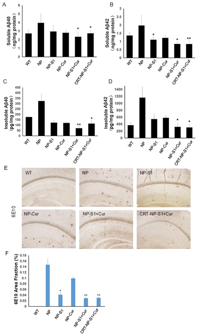
The levels of soluble Aβ40 (A) and Aβ42 (B), insoluble Aβ40 (C) and Aβ42 (D) in the brain lysates of AD mice treated with NP control, NP-S1, NP-Cur, NP-S1+Cur and CRT-NP-S1+Cur were detected by Aβ40 and Aβ42 sandwich ELISA kits, respectively. The senile plaques in the brains of AD transgenic mice treated with NP control, NP-S1, NP-Cur, NP-S1+Cur and CRT-NP-S1+Cur were detected by immunohistochemistry (E) and quantitatively analyzed by Image-Pro Plus software (F) (*, P < 0.05, **, P < 0.01, compared with NP control-treated AD mice).