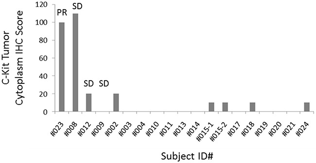
Figure 4. Baseline cytoplasmic c-Kit tumor IHC score.

c-Kit staining was quantified by using a 4-value intensity score (none = 0; weak = 1+; moderate = 2+; strong = 3+) and the percentage (0%-100%) of the extent of reactivity. A final score was obtained by multiplying the intensity and reactivity extent values (range, 0-300). Subject 023 (PR of 151 days) and subject 008 (SD of 256 days) showed the highest H-score. (Archival FFPE tissue was available in 18 subjects. c-Kit was not detected by IHC in 11 subjects).
