
Figure 2. Axial T1-weighted contrast enhanced (top two rows) and T2-FLAIR images (bottom row) in Patient 6 with an original left temporal glioblastoma as depicted in the middle row.
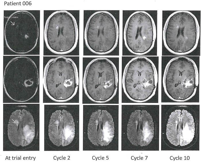
The patient developed a right frontal periventricular tumor progression, marked with the arrow as the index tumor at baseline. Follow-up images showed stable disease of the index tumor and as for the left temporal tumor component evolving multi-cystic contrast-enhancing changes and edema were noted.