
Figure 3. Axial T1-weighted contrast-enhanced images (top row) in Patient 4 with right temporal glioblastoma at trial entry with corresponding T2-FLAIR (middle row) and corresponding DWI with apparent diffusion coefficient (ADC) sequences (bottom row).
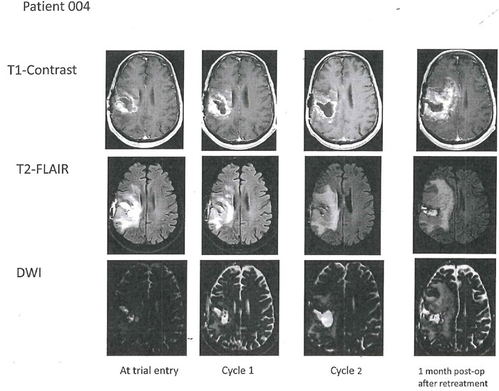
After two cycles with AXL1717 there was enlargement of the tumor cavity. Surgical resection histopathology showed abundant necrosis with small islands of viable tumor. Progressive disease on imaging (and clinically) after an additional treatment cycle (post-operative; last column) and the patient was removed from the trial. T2 FLAIR shows increased edema post-operatively one month after treatment.