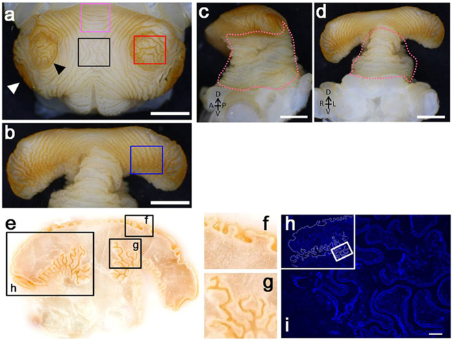
Figure 2.
Complex furrows on the surface of the horn primordial. (a to d) Photos of a completely formed horn primordia dissected from larval (prepupal) head. (white scale bars indicate 2 mm) (a) Top of the cap region. Two concentric circles can be clearly recognized. (b) Underside of the cap region. (c and d) Side view and frontal view of the horn primordia, respectively. Lifting the cap of the horn primordia exposes the accordion-like folding pattern of the stalk (pink dotted lines). (e) Cryostat frontal section of a fully developed horn primordia: depth and pattern of the furrows differ among different regions. (f) Furrows at the top of the cap region are less deep, compared to furrows in other regions. (g) Many deep and branched furrows can be recognized at the underside of the cap. (h) Hoechst stained section of a horn primordia (region corresponds to window in (e). The border of the horn primordia is artificially outlined as a white line. (i) Magnified image of the region depicted by a window in (h). Cells are aligned along the surface of the furrows. No solid tissue is present inside the primordia (scale bar indicates 100 µm).

