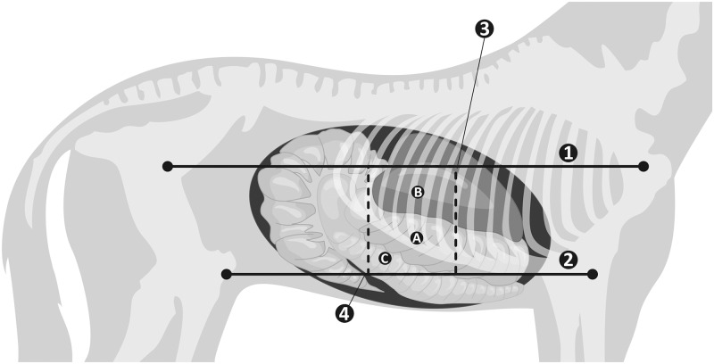
Fig 1.
Schematic illustration of the right side of the abdomen for the examination of the right dorsal colon—RDC (A). The imaging window was allotted using four lines: line 1 –from the shoulder joint to the coxal tuberosity, line 2 (parallel to line 1)–from the elbow joint to the stifle joint, line 3 –a line along the 9th intercostal space, line 4 (parallel to line 3)–a line along the 15th intercostal space. The liver was visible next to the RDC (B), the right ventral colon was visible below the RDC (C).

