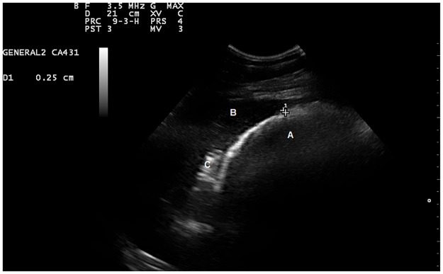
Fig 2. A normal sonogram of the right dorsal colon in a healthy pony.
Image obtained by placing a convex transducer in the right 12th intercostal space. The right dorsal colon (A) was identified as a semicircle with bowel gas beam reflections. The liver (B) is seen in the upper hand corner of the image, the duodenum (C) is seen between the liver and the right dorsal colon. The measured thickness of the colonic wall was 0.25 cm.

