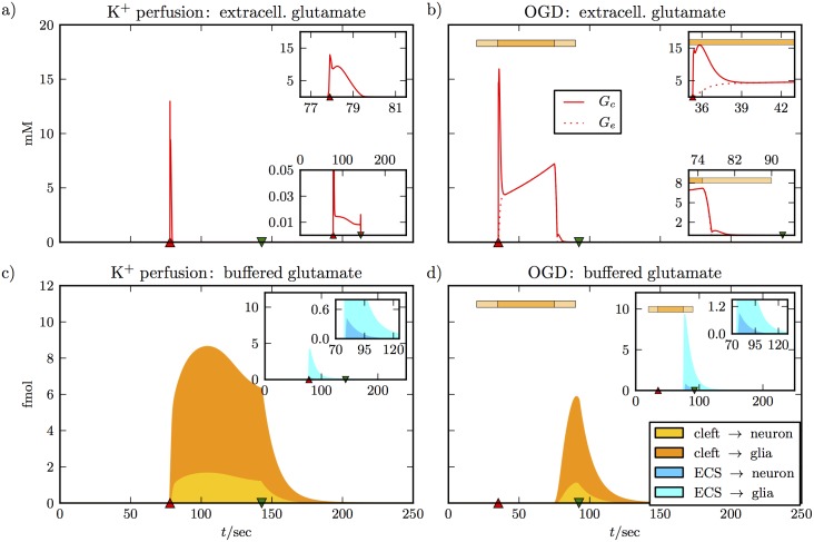
Fig 3. Glutamate dynamics for the simulations from Fig 2.
Panels (a) and (b) show a jump of the cleft glutamate concentration to about 15 mM when the neuron depolarizes. In the simulation of K+ perfusion we assume intact glutamate uptake at all times, and the high cleft concentration is brought back to a much lower level within 2 sec (see upper inset of (a)). There is a plateau concentration near 0.01 mM that is maintained as long as the neuron is depolarized, and is only cleared after repolarization (lower inset of (a)). There is no noticeable glutamate elevation in the ECS. In panel (b) we see different dynamics, because there is no glutamate clearance during OGD. The sharp jump in the cleft concentration also decays within 2 sec, but the concentration then settles to a much higher level of 5 mM (see upper inset). It keeps increasing slowly until glutamate clearance is slowly reactivated (see horizontal bar for the OGD protocol). The lower inset shows that glutamate is back to a very low level before the neuron repolarizes. The extracellular concentration goes up to more than 5 mM. When there is no glutamate clearance at all (center part of the OGD bar) the concentrations in the cleft and the ECS are equal because of diffusion. The lower panels show pathways of glutamate clearance. Glutamate that has been taken up from the cleft or the ECS by either the neuron or the glial cell, but has not yet been recycled, counts as buffered glutamate (see Eq 30). The main plots show glutamate clearance from the cleft, the inset show clearance from the ECS. For K+ perfusion, more glutamate is cleared directly from the cleft than from the ECS (see peak values in the main plot and inset of panel (c)). In the cleft, more glutamate is cleared by glia than by the neuron (compare the orange and the yellow portion of the total uptake from the cleft). In the ECS, this relation is even more pronounced (compare turquoise to blue in the second inset). In panel (d), glutamate clearance only sets in with the reactivation of regulatory functions at the end of the OGD protocol. Now more glutamate is cleared from the ECS, since there is more in the ECS than in the clefts. The relation between uptake by glia and neural uptake is consistent with (c) and Eqs (26) and (18).

