Figure 1.
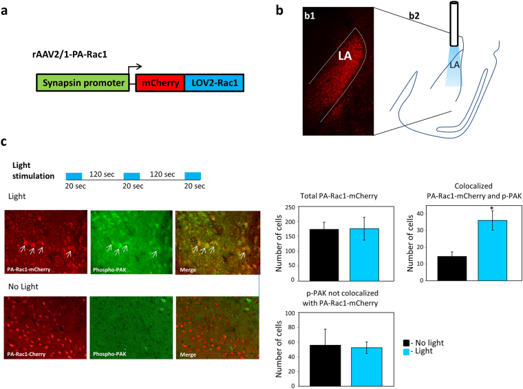
Activation of Rac1 GTPase in LA leads to increase in PAK phosphorylation. (a) Schematic description of the vector expressing Rac1 conjugated to LOV2 (PA-Rac1) and mCherry under the control of synapsin1 promoter. (b) PA-Rac1 is expressed in lateral amygdala (LA) as depicted by expression of mCherry (b1). The area of illumination by optic fiber includes the LA (b2). (c) Phospho-PAK is measured in the amygdala in light stimulated animals 30 minutes after stimulation (same light stimulating protocol used in behavior) and in animals that were not subjected to light. Left panels are representative immunohistochemistry of phospho-PAK and PA-Rac1-mCherry expression in AAV injected animals in light and no light animals. Arrows point at examples of phospho-PAK in PA-Rac1 expressing cells. Right graphs are quantifications of labeled cells (n = 6 mice no light, n = 7 light). The mean of total number of cells from slices from all animals in each group is shown. More phospho-PAK is detected in cells expressing PA-Rac (mCherry) in light exposed animals compared with animals that were not subjected to light (p < 0.02).

