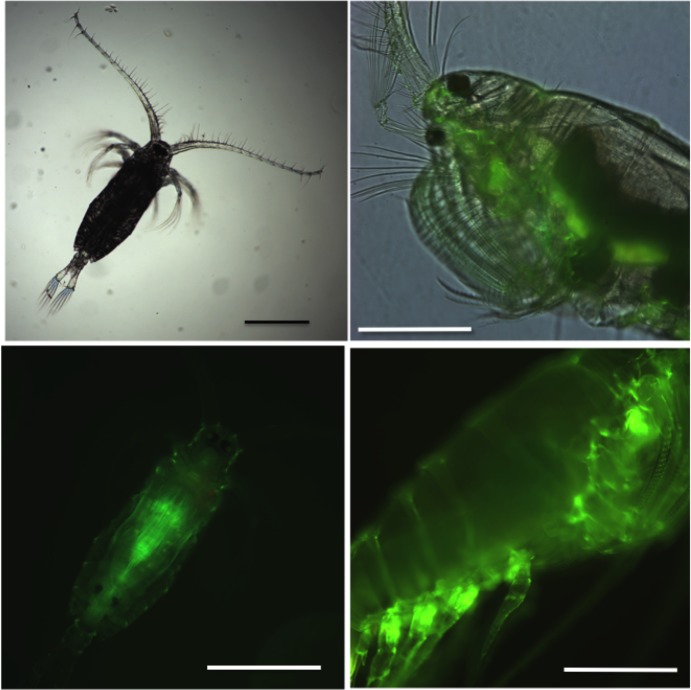
Fig 1.
Light micrographs of Labidocera madurae copepodite (A, B) and adult female (C,D). (A) Copepodite stage CIII, dorsal view (magnification: 4x). (B) Same copepodite as in A under fluorescent light showing expression of green fluorescent protein (GFP) (magnification 10x). (C) lateral view of the anterior portion of an adult female showing one dorsal and the ventral ocelli, feeding appendages and GFP expression (magnification 10x). (D) Lateral view of the same individual as in C under fluorescent light showing GFP expression at the base of the swimming legs (magnification 10x). Scale bar: 0.5 mm.

