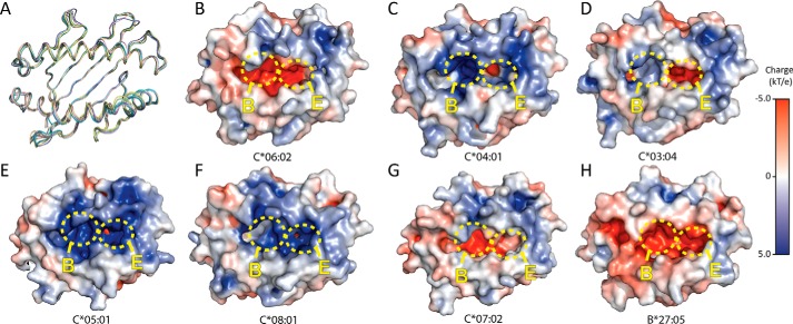
Figure 6.
Comparison of available HLA-C structures. A, Cα trace overlay of HLA-C*06:02 (teal), HLA-C*03:04 (PDB 1EFX) (green), HLA-C*04:01 (PDB 1QQD) (pink), HLA-C*05:01 (PDB 5VGD) (purple), HLA-C*07:02 (PDB 5VGE) (yellow), HLA-C*08:01 (PDB 4NT6) (wheat) and HLA-B*27:05 (PDB 3BP4) (blue). B–H, surface electrostatics of: HLA-C*06:02 ARTE (B), HLA-C*04:01 (C), HLA-C*03:04 (D), HLA-C*05:01 (E), HLA-C*08:01 (F), HLA-C*07:02 (G), and HLA-B*27:05 (H). B- and E-pockets are shown as yellow circles. Structures were generated using APBS Tools plug-in within PyMOL.

