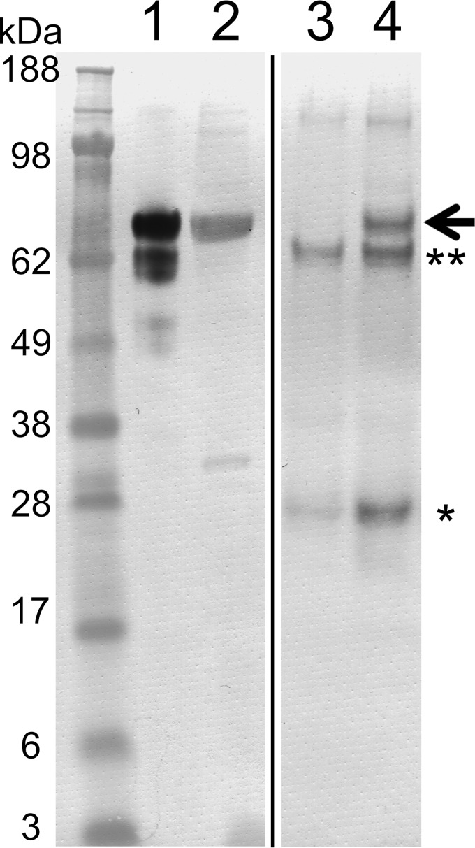
Figure 1.
Western blot analysis using an anti-FKBP65 antibody on proteins immunoprecipitated from chicken rER extracts by an Hsp47 antibody. Proteins immunoprecipitated from chicken rER extracts using a monoclonal Hsp47 antibody were electrophoresed on a Novex NuPAGE Bis-Tris 4–12% gel under reducing conditions. Proteins were transferred to a PVDF membrane and subsequently analyzed by Western blotting using a polyclonal FKBP65 antibody. Lane 1, chicken rER extract (immunoprecipitate input); lane 2, purified chicken FKBP65; lane 3, immunoprecipitate using protein G-Sepharose alone; lane 4, immunoprecipitate using protein G-Sepharose plus Hsp47 mAb. The arrow highlights the position of FKBP65. * and ** are protein G and preincubated BSA bands from the protein G-Sepharose. The black line spacing denotes irrelevant lanes that were eliminated from the image.

