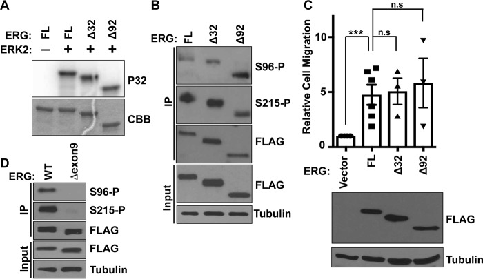
Figure 4.
Comparison of common TMPRSS2-ERG fusion products and alternative splicing isoforms. A, in vitro phosphorylation of full-length (FL) and the indicated N-terminal deletions of ERG by ERK2. Coomassie (CBB) staining of gel (bottom panel) and autoradiograph of 32P-labeled (P32) ERG (top panel) are shown. B, immunoblot using Ser(P)-96 or Ser(P)-215 specific antibodies or FLAG antibody of whole cell extract (input), protein immunoprecipitated (IP) with FLAG antibody from RWPE1 cells expressing 3×FLAG-ERG (FL), N-terminal deletions 3×FLAG-Δ32ERG (Δ32), or 3×FLAG Δ92ERG (Δ92). C, Transwell migration of RWPE1 cells stably expressing indicated 3×FLAG-ERG proteins, shown relative to RWPE1 cells with empty vector as mean and S.E. (n = 3). p values were determined by t test: *, p < 0.05; **, p < 0.01; ***, p < 0.001. Expression shown by immunoblot. D, FLAG IP immunoblot with indicated antibodies from RWPE1 cells expressing ERG or ERG lacking exon 9.

