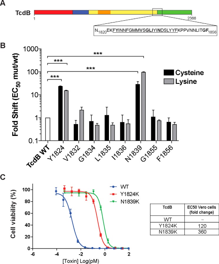
Figure 1.
Identification of functional defects at the boundary of the CROPs and translocation domain. A, schematic drawing of TcdB organized into four domains: N-terminal glucosyltransferase domain (red), autoprotease domain (blue), translocation/pore formation domain (yellow), with the hydrophobic region (residues 956–1128) shown in orange, and the C-terminal CROPs repeats domain (green). The box represents the hydrophobic region at the junction under investigation (N1820-F1856). B, effects of Cys and Lys substitutions on cellular toxicity. Viability of each Cys and Lys mutant was tested by exposing titrated mutant soluble lysates onto CHO-K1 cells (3-fold titration, 3 days) and quantified using the PrestoBlue fluorescence viability assay. Fold shift of mutant to wild type was calculated by dividing the half-maximum effective concentration (EC50) of mutants by the EC50 of wild type. n = 3. ***, p < 0.001, two-way analysis of variance. C, toxicity of mutant and wild type toxins on Vero cells was quantified by titrating purified proteins onto Vero cells (4-fold titration) in 96-well plates and incubated for 24 h at 37 °C (n = 3). Forty-eight hours later, the cell viability of treated cells was quantitated by SRB staining.

